Methods for extracting volatiles
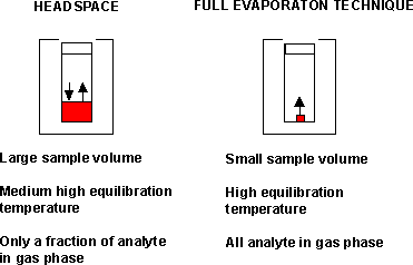 As related to the vial volume, the amount of sample
that can be used for extraction by headspace is about 10%, and by the full
evaporation technique about 0.15% . Because headspace extraction allows for the
use of a large sample size, it gives a higher sensitivity than the full
evaporation technique, and is used for the screening process.
As related to the vial volume, the amount of sample
that can be used for extraction by headspace is about 10%, and by the full
evaporation technique about 0.15% . Because headspace extraction allows for the
use of a large sample size, it gives a higher sensitivity than the full
evaporation technique, and is used for the screening process.
In contrast to headspace, however, the full evaporation technique favours a total release of the analyte into the gas phase. Under such a condition the matrix effect will be negated, making the full evaporation technique suitable for quantitative analysis.
Back to previous text or contents , or forwards to apparatus set up in next analysis step.
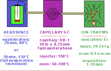 Apparatus set up for analysing volatiles
The extraction is done by static headspace for the screening and by the full evaporation technique for the quantitation. The chromatographic separation is done in a capillary with an apolar stationary phase. This is to make the screening and also the search in the literature for retention indices matching the analyte´s retention index as versatile as possible. To promote a high sensitivity and broad range of selectivity, the detection is done by an ion-trap with the mass spectrometer run in the scan mode.
Back to previous text or contents , or forwards to analyte detection in next analysis step.
Search for the volatile "general unknown"
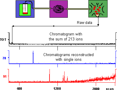 The
results generated by the apparatus set up may be regarded only as raw data that
need refinement before a final analytical assessment can be done. The method of
the first main step to reach this goal is shown here in the figure taken from
the analysis on a blood sample from a fire victim of an accidental fire.
Because of the high background level generated by the 213 ions, the total ion
current of the top chromatogram (in black) reveals no peaks.
The
results generated by the apparatus set up may be regarded only as raw data that
need refinement before a final analytical assessment can be done. The method of
the first main step to reach this goal is shown here in the figure taken from
the analysis on a blood sample from a fire victim of an accidental fire.
Because of the high background level generated by the 213 ions, the total ion
current of the top chromatogram (in black) reveals no peaks.
To be able to detect trace amounts of the volatile "general unknown" the background noise must be diminished. This is done by reconstruction of the total ion current with each of the 213 ions monitored. Even though such a process involves only computer work and no "wet chemistry", it may be a tedious proceeding taking several hours when carried out manually, but by using a macro program the reconstruction process is facilitated and speeded up.
As seen in the figure, when the total ion current is reconstructed with the m/z 78 ion (blue chromatogram), one clear peak shows up, and when the ion with the m/z 91 is used (red chromatogram) four peaks appear. To be able to judge whether a peak is a part of the background noise or arises from an analyte, the limit of detection, i.e. a signal response equal to three times the standard deviation of the gross blank signal, is measured according to Knoll JE: Estimation of the limit of detection in chromatography; J Chromatogr Sci 23: 422; 1985.
Back to previous text or contents , or forwards to analyte identification in next analysis step.
Identification of detected volatile "general unknown"
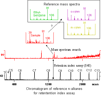 The second main step in the
process of handling the raw data involves the identification of a detected
volatile "general unknown". To show the technique used to reach this
goal, I have chosen as an example the second peak from the left on the
chromatogram (in red) reconstructed with the ion at m/z 91. By searching
an on-line library for mass spectra that best fit the spectrum of the
"general unknown" (in red), a number of candidate substances are then
first picked. Since the hydrocarbons within a series often yield very similar spectra,
it is usually impossible to classify such an analyte based only on its mass
spectrum. Also, in the example shown in the figure, the sample may be
identified as any of the candidates, ethylbenzene (green), o-xylene (violet),
or m-xylene (yellow).
The second main step in the
process of handling the raw data involves the identification of a detected
volatile "general unknown". To show the technique used to reach this
goal, I have chosen as an example the second peak from the left on the
chromatogram (in red) reconstructed with the ion at m/z 91. By searching
an on-line library for mass spectra that best fit the spectrum of the
"general unknown" (in red), a number of candidate substances are then
first picked. Since the hydrocarbons within a series often yield very similar spectra,
it is usually impossible to classify such an analyte based only on its mass
spectrum. Also, in the example shown in the figure, the sample may be
identified as any of the candidates, ethylbenzene (green), o-xylene (violet),
or m-xylene (yellow).
An additional identification criterion is, thus, needed. To this end, the candidates' retention index values are measured and compared with those as found in the literature or in a retention index library. These three-digit integers denote the retention time of a compound relative to the retention times of homologous n-alkanes (chromatogram in black) as indicated in the figure. Since the unknown analyte in the example shows up between the n-alkanes with eight and nine carbon atoms, it has the retention index value of 840, and was identified in the example as ethylbenzene (green).
The retention indices may, in contrast to the retention times, be transferable between different gas chromatography systems. This situation opens up access to comparable data in a retention index library or in a vast number of reports in the literature, thus, making the analyst less dependent on the list of his own reference substances.
Back to previous text or contents, or see table for summary of analytical procedure.
|
|
||
|
STEP BY STEP PROCESSING ION-TRAP RAW DATA |
||
|
Detection |
Identification |
Quantitation |
|
By the use of a data program under "DATAMASTER", reconstitute 213 chromatograms with each monitored ion of the total ion current To reach optimal relation between signal response of unknown analyte and of base-line noise, fine-tune reconstituted chromatograms by adding up 2 or 3 ions Control that unknown analyte´s peak height exceeds 3 times the standard deviation of the base line noise |
Search on-line library for analyte´s mass spectrum On the basis of the mass spectra found, pick possible candidates and notify their CAS numbers Search literature for candidates´ retention indices On the basis of the retention times of the n-alkanes series (butane to tridecane), calculate analyte´s retention index Compare data from analysis of candidates´ mass spectra and retention indices |
Control that unknown analyte´s peak height exceeds 10 times the standard deviation of the base line noise Extract test sample by the headspace variant, "full evaporation technique", i.e. equilibrate a small sample size at a high temperature Construct calibration graph for "full evaporation technique" |
Back to previous text , or to contents